Learn more about Tissues
In this module, you will learn more about
- What is a tissue?
- Epithelial tissues
- Connective tissues
- Muscle tissues
- Nervous tissues
Learn even more: understanding tissues is a foundation for understanding all the organs covered in your other learning modules
1 What is a tissue?
In humans, cells of the same type, or shared broad functions, are organised into a tissue.
The consequence of this is that the properties and functions of any tissue are the result of the particular cells that comprise it.
For example the alveoli in the lung are made up of a tissue called simple squamous epithelium which is a single sheet of very thin cells. This is ideally suited for the exchange of gases between the alveolar air and capillary blood.
In this module, we will go through the four major tissue types and in each case highlight important sub-types.
2 Epithelial tissues
https://commons.wikimedia.org/wiki/File:Illu_epithelium.jpg
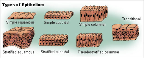
What characterises all epithelial tissues is that they are composed of closely packed cells. You can see that in the diagram directly above.
There are two ways we classify epithelial tissue
There are two ways we classify epithelial tissue
- What is the shape of the predominant cell type?
- How many layers are there?
Classification by shape
When classified by shape, three types of epithelium are identified:
When classified by shape, three types of epithelium are identified:
- squamous: flattened cells
- cuboidal: the cells are generally as high as they are wide (like a cube)
- columnar: the cells are taller than they are wide (like a column)
Classification by number of layers
When classified by number of layers, two types are identified:
Whether an epithelium is simple or stratified the basal surface of the tissue will essentially be “glued” to the underlying tissue by a layer of extracellular material called the basement membrane.
When classified by number of layers, two types are identified:
- simple: a single layer of cells
- stratified: multiple layers of cells
Whether an epithelium is simple or stratified the basal surface of the tissue will essentially be “glued” to the underlying tissue by a layer of extracellular material called the basement membrane.
Epithelial cells: special cases
(i) The lining the trachea and the larger airways the epithelium is of the pseudo-stratified columnar type. It is called thus because unlike what is normally seen the nuclei of adjacent cells are at different positions, giving the illusion that the tissue is stratified
(ii) In regions like the bladder the inner lining is a transitional epithelium. This is a stratified epithelium but the cells have a unique property of being able to become flatter as the organ fills with urine
(iii) Stratified epithelium is most commonly of squamous cells (although generally the cells at the most basal layer are cuboidal). In a case like the epidermis of the skin, aside from becoming increasingly flattened: the cells of the upper most layer undergo other changes. The most significant change being the cytoplasm is largely replaced by the protein keratin. This gives rise to so called keratinised epithelium.
3 Connective tissues
The most distinguishing thing about all connective tissues is that in all cases (bar one) much of the tissue is extracellular material commonly an assemblage of water, salts, other low molecular substances and proteins. This assemblage is called the extracellular matrix and the cells of the connective tissue are embedded or immersed in this matrix.
This common feature actually allows for a number of structurally very distinctive tissues to all be classified as connective tissue
This common feature actually allows for a number of structurally very distinctive tissues to all be classified as connective tissue
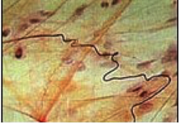
Loose or areolar connective tissue
https://upload.wikimedia.org/wikipedia/commons/4/47/Illu_connective_tissues_1.jpg
Areola means “an open space”. In this tissue, the fibres and cells are in a loose arrangement surrounded by what (under the microscope) appears to be empty space. The space is actually filled by the extra-cellular matrix. In the diagram you can see the cells (the darker nuclei being the most obvious feature) are widely spaced. Strands of extracellular protein also visible
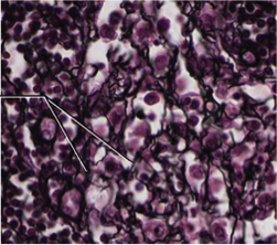
Reticular tissue
https://commons.wikimedia.org/wiki/File:410_Reticular_Tissue.jpg
“Reticular” in general relates to a network. In connective tissue of the reticular type the fibres are arranged in a mesh. In a number of organs (including the lymph nodes, spleen and bone marrow) the reticular connective tissue forms the framework, the stroma, of these organs with the functional tissue, the parenchyma interspersed through the stroma.
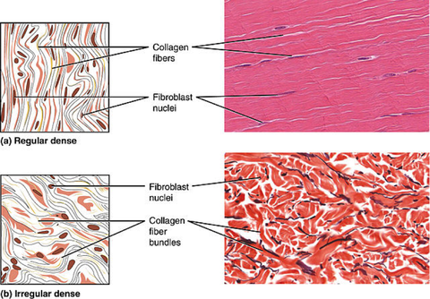
Dense connective tissue
https://commons.wikimedia.org/wiki/File:411_Reg_Dense-Irregular_Dense.jpg
This tissue is dense because so much of it is made up of fibres of the protein collagen. In the images above you can see the cells of this tissue (the fibroblasts) are essentially “wedged” in between strands and clumps of fibres. You can also see that sometimes there is an orderly arrangement of the fibres and sometimes not so much. The predominance of fibres in this tissue (c.f. with areolar and reticular connective tissues) gives dense connective tissue greater resistance to being stretched out of shape (“tensile strength”)
Adipose tissue

https://commons.wikimedia.org/wiki/File:Yellow_adipose_tissue_in_paraffin_section_-_lipids_washed_out.jpg
Adipose tissue is unique among the connective tissue types in that the bulk of the tissue is made up of cells: adipocytes. In the picture about each of the big white polygons is an adipocyte and in these cells the bulk of the cytoplasm is lipid.
Cartilage
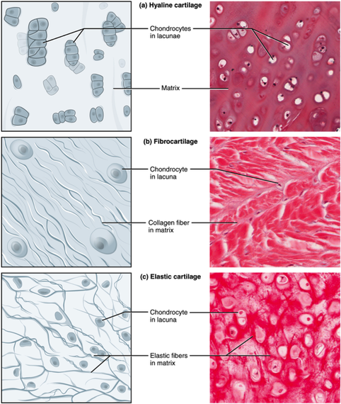
https://commons.wikimedia.org/wiki/File:412_Types_of_Cartilage-new.jpg
Compared to the connective tissues described in the previous sections, cartilage is more firm and as a result it provides in some cases structural support (as in the nose and ears and costal cartilage) or resistance to compressive stress (as in the discs between the vertebra of the spinal column). The cells found in mature cartilage are called chondrocytes. (Interesting bit of medical information: that is why the two abdominal regions lateral to the epigastric are called hypochondriac: they lie below the costal cartilage). Differences in the composition and properties of the extracellular matrix result in the three sub-types of cartilage shown in the figure above
- hyaline (“glassy”) cartilage is firm and very smooth.
- in elastic cartilage there is a predominance of elastic fibres
- in fibrocartilage the fibres are coarse and densely packed
Two other types of connective tissue
Blood and bone are indeed connective tissues and that’s a reasonable claim since in both you have cells surrounded by a matrix. In the blood, the matrix is a fluid: plasma. In bone the matrix is very hard because of heavy deposits of mineral (including calcium and phosphorus) salts.
Blood and bone are indeed connective tissues and that’s a reasonable claim since in both you have cells surrounded by a matrix. In the blood, the matrix is a fluid: plasma. In bone the matrix is very hard because of heavy deposits of mineral (including calcium and phosphorus) salts.
4 Muscle tissues
Depositphotos.com
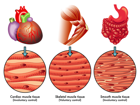
Like epithelial tissue (and unlike most types of connective tissue), the bulk of muscle tissue is made up of cells.
The cells of muscle are called fibres because of their shape and how they are arranged in muscles. What distinguished muscle fibres from the cells of the other tissues described so far is that muscle fibres are “excitable” (that is they can produce an electric signal known as an action potential) and capable of contraction (that is reducing in length).
There are three types of muscle tissue which differ in the features of the muscle fibres and how fibres are arranged. Here is a table that lists the differences.
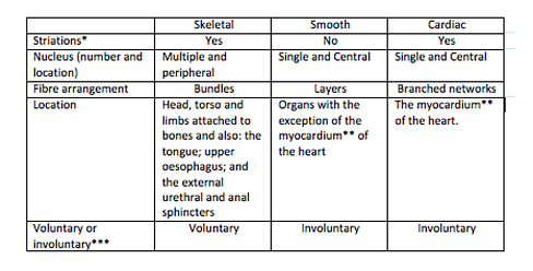
* Striations are alternating bands of light and dark which can be seen in skeletal and cardiac muscled fibres when examined under a microscope. They are the result of the highly ordered arrangement of the thin strands of protein (myofilaments) that make up the bulk of the cytoplasm of the fibre and are responsible for fibre contraction
** This is the major layer of the walls of the chambers of the heart
*** Voluntary muscle requires signals from the somatic motor system for contraction. In contrast involuntary muscle is under the control of the autonomic nervous system but may also include fibres that generate their own signal for contraction (so called “pacemaker” cells). The distinction can be thought of this way
- You can move your limbs, head and torso and you control expulsion of urine and faeces
- You can’t command the movement of food through the gastro-intestinal tract or how quickly your heart beats or how much blood flows to your skin at any one time.
5 Nervous tissues
As the name indicates this tissue is found in the brain, spinal cord and peripheral nerves. It is highly cellular comprising two distinctive cells types:
- Neurons: These are excitable cells, that is these cells are capable of generating an action potential
- Glia or neuroglia: These cells provide support to neurons