Learn more about the Nervous System
In this module, you will learn more about
- The nervous system overall
- The central nervous system (CNS)
- The peripheral nervous system (PNS)
- The reflex arc
- Pain
Learn even more: See Chemistry and Cells modules
1 The nervous system overall
Organisation of the nervous system
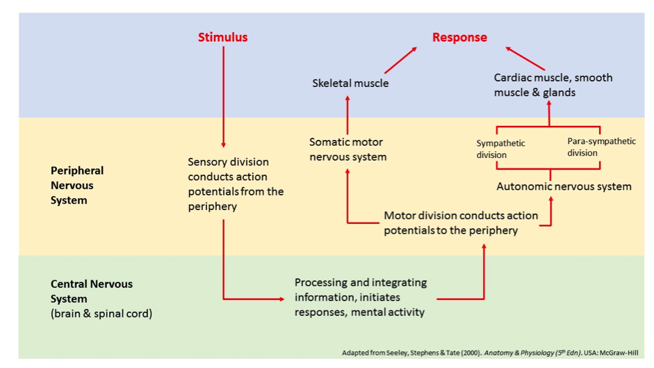
The nervous system has two major anatomical subdivisions, the Central Nervous System (CNS) and the Peripheral Nervous System (PNS). The PNS is further subdivided as you can see above.
The neuron
|
|
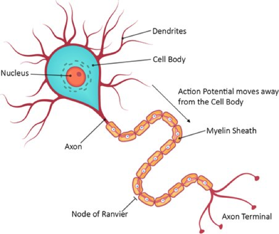
The Structure of a Neuron (Depositphotos.com)
|
The structural unit of the nervous system is the neuron. These cells carry electrical/chemical messages to and from the CNS. They are helped in their job by 5 different kinds of supporting cells known as glial cells. Sensory neurons carry input from the body or the external environment to the CNS while motor neurons carry information from the CNS to muscles or glands. Neurons of the autonomic nervous system control organs in the body such as the gut and endocrine glands.
A neuron uses changes in its membrane potential to transmit electrical signals to and from the CNS. The concentration of sodium is higher outside the cell and that of potassium is higher inside the cell. Sodium therefore, wants to move down its concentration gradient into the cell and potassium wants to move down its concentration gradient out of the cell. Indeed, the cell membrane is permeable to both these ions, which leak in or out of the cell. The sodium-potassium ATPase pump counteracts this movement by pumping sodium out and potassium into the cell, maintaining a membrane potential of -70 mV. Increases in this membrane potential create action potentials, which are the electrical signals used to transmit information along the neuron.
A neuron uses changes in its membrane potential to transmit electrical signals to and from the CNS. The concentration of sodium is higher outside the cell and that of potassium is higher inside the cell. Sodium therefore, wants to move down its concentration gradient into the cell and potassium wants to move down its concentration gradient out of the cell. Indeed, the cell membrane is permeable to both these ions, which leak in or out of the cell. The sodium-potassium ATPase pump counteracts this movement by pumping sodium out and potassium into the cell, maintaining a membrane potential of -70 mV. Increases in this membrane potential create action potentials, which are the electrical signals used to transmit information along the neuron.
Signal transduction
Signal transduction is the process the body uses to turn complex phenomena like light, sound waves, taste, smells (volatile chemicals in the nasal passage) and even chemical messengers into messages that can be sent around the body, to and from the brain. These messages are processed (mostly) in the brain and spinal cord. Specialised receptors collect incoming information (photons of light, chemicals and so on) and change it into electrical messages that are sent through neurons to and from the central nervous system.
Neurotransmitters
|
|
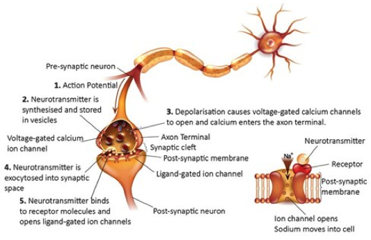
The Synapse (Depositphotos.com)
|
Neurotransmitters are chemicals, usually made in neurons, which enable transmission of messages. These chemicals are released at the ends of neurons into the synaptic cleft (space between neurons in a neuron chain or between neurons and end effector organs). Electrical messages travel along the neuron and trigger an influx of (typically) calcium ions into the axon terminals. The influx of calcium enables neurotransmitter molecules stored in vesicles (membrane-bound storage containers) inside the axonal terminal to be pulled towards the end of the neuron, to the synaptic cleft. Once the vesicles are at the edge of the neuron the neurotransmitter moves into the space outside the neuron via exocytosis. The neurotransmitter diffuses across the synapse and ‘binds’ to receptors on the post synaptic space (‘after the synapse’ space) where they enable the opening of chemically gated ion channels, thus allowing the transmission of the action potential to continue.
2 The central nervous system (CNS)
The central nervous system consists of the brain and spinal cord. It is like a massively complex computer chip, collecting information from sensors inside and outside the body and processing the information so it can send out ‘action’ messages. Some neurons in the CNS are ‘insulated’ and so send messages superfast, while others are slower but have more surface area to collect incoming information. The central nervous system is so important it is floated and protected by cerebrospinal fluid (CSF) and is wrapped in layers of tissue called the meninges.
The spinal cord
The spinal cord is a two-way system that sends messages to the brain and relays responses back to the body. Stretching from the foramen magnum of the skull to the first or second lumbar vertebrae, the spinal cord is protected by bone, dura mater, an epidural space containing fat, and cerebrospinal fluid contained within the subarachnoid space. Grey matter is located in the centre of the spinal cord and white matter on the outside.
There are 31 pairs of spinal nerves (clusters of neurons), which leave the vertebral column via intervertebral foramen and travel to their respective body region. Reflex actions are initiated and completed at the spinal cord (see the section on the reflex arc).
There are 31 pairs of spinal nerves (clusters of neurons), which leave the vertebral column via intervertebral foramen and travel to their respective body region. Reflex actions are initiated and completed at the spinal cord (see the section on the reflex arc).
The brain
|
|
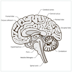
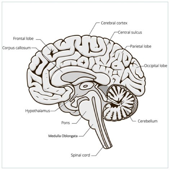
The Structure of the Brain (Depositphotos.com)
|
The brain is composed of fragile nervous tissue which is protected by the skull on the external surface and several layers of tissue. The meninges form three membranous layers, with the pia mater being closest to the brain, a middle layer known as the arachnoid layer and an outer layer of dura mater. The subarachnoid space, lying between the pia mater and the arachnoid layer, contains cerebrospinal fluid (CSF) which cushions the brain, protects it from trauma, and nourishes it.
The cerebral hemispheres form the superior part of the brain and account for about 83% of the brain’s mass. Visually, elevated ridges of brain tissue known as gyri can be seen with shallow grooves known as sulci separating them. Deep fissures separate the larger regions of the brain. The right and left hemispheres of the brain are separated into five lobes; frontal, parietal, temporal, occipital and insula. The cerebral hemispheres are separated from the inferior cerebellum by a transverse cerebral fissure.
The cerebral cortex is where the conscious mind is found, allowing us to be conscious of the world around us, to communicate, to remember and to move. It contains billions of neurons and has three functional areas; motor, sensory and association areas. Being the end site and primary processing centre for sensory information, different sorts of information go to different parts of the brain surface, in the cortex, in a highly organised and very typical pattern. For example, most information that comes through the eyes (visual information) is sent to the visual cortex, which is located in the occipital lobes (at the back of the brain). Some say this is why a hit on the back of your head gives you the impression that you are ‘seeing stars’. Some visual ‘memories’ are also stored in the occipital lobes, helping you to remember things you have seen before.
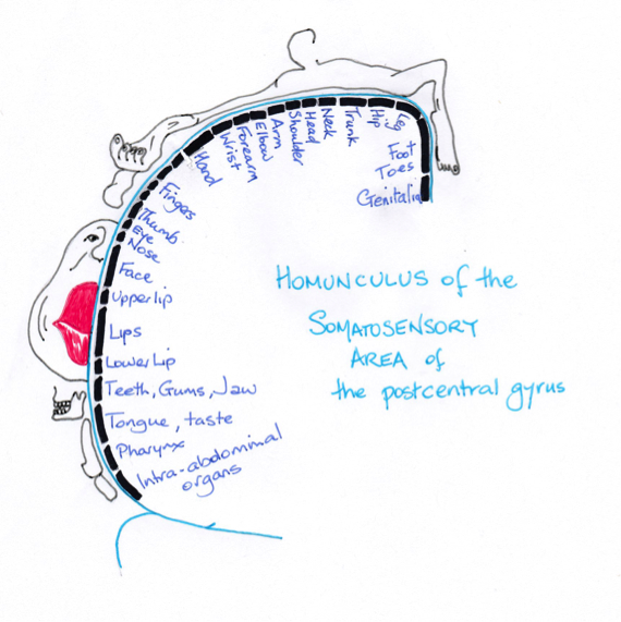
© Dr Patricia Logan 2017: © Dr Patricia Logan 2017: Homunculus of the somatosensory area of the post central gyrus, adapted from McNaught & Callander, 1997
The primary somatosensory cortex is located in the post-central gyrus (the area posterior to the central sulcus) of the parietal lobe. Neurons receives sensory information from sensory receptors in the skin, muscles, tendons and joints, and are able to identify the part of the body being stimulated (spatial discrimination), in a highly systematic manner. Neurons carrying sensory information from specific parts of the body terminate in the somatosensory cortex where information is represented in the form of a ‘body map' (somatosensory homunculus), which forms a spatial, upside down image of the body. It is because of this map that we are able to localise sensations (for example, we know we just banged our right index finger on the computer key – not our left nostril). The amount of space allocated to sections of our body on the map is distorted and not related to the size of that section of body, but rather to the amount of sensory information coming from that section of body; the face especially the lips, the genitals and the fingers and toes, are disproportionately large, due to their acute sensitivity.
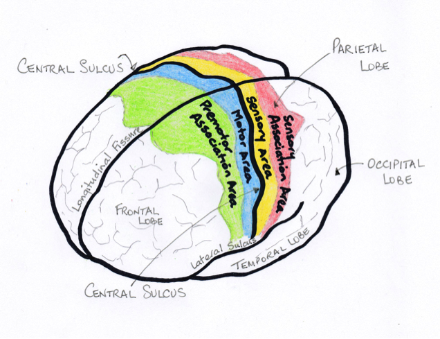
© Dr Patricia Logan 2017: Somatosensory association cortex, adapted from McNaught & Callander, 1997
Posterior to the somatosensory cortex and connecting to it, lies the somatosensory association cortex. The primary function of this area is to integrate incoming information such as temperature and pressure from the primary somatosensory cortex, in order that one can make sense of the information, such as the shape, size and texture of an object.
Sitting right next to the somatosensory cortex, located in the precentral gyrus of each frontal lobe lies the primary motor cortex. Like the somatosensory cortex, it has a motor homunculus that relates to parts of our body. Large pyramidal cells allow conscious and precise control of voluntary movements of skeletal muscles. An important thing to remember is that both motor and sensory nerve pathways crossover (decussate or go from left side to right side) before they get to higher brain centres, so that the right motor cortex controls the left side of the body and sensory information from the left side (e.g. left hand) is mapped onto the right somatosensory cortex.
The premotor cortex lies anterior to the precentral gyrus and is involved in planning movement. This region co-ordinates movement of several muscle groups and is responsible for skilled motor activities such as playing a musical instrument.
The Structure of the Brain (Depositphotos.com)
On both sides of the brain are the temporal lobes which collect and collate auditory (sound) information. Areas in these parts of the brain are important for sound-related understanding, such as the understanding of language. A common brain injury resulting from a stroke is damage to one part of the temporal lobe (Wernicke’s area) on the left side of the brain. This damage means a person has difficulty understanding (perhaps remembering) spoken language.
The diencephalon consists of the thalamus, the hypothalamus and the epithalamus, and is surrounded by the cerebral hemispheres. The thalamus receives afferent information sorts it and sends it to the respective region of the cerebral cortex, it helps regulates emotion, directs activities of the motor cortex, and is involved in memory and sensory integration. The hypothalamus is the main control centre for visceral organs and for the control of homeostasis.
Perhaps the most important brain area for maintaining basic life functions is the brain stem, consisting of the pons and medulla oblongata. The brain stem is located at the base of the brain at the top of the spinal cord. It is the built in ‘nurse’ continually monitoring vital signs such as heart and respiratory rate, blood pressure, body fluid components and so on. It helps adjust these vital signs to ensure the body maintains physiological homeostasis.
The ‘lower’, some say ‘more primitive’ parts of the brain assist and support with complex information transmission and integration. Areas such as the midbrain, located between the diencephalon and the pons, support integration of sensory and motor information in complex patterns. People with diseases like Parkinson’s disease, with its typical tremor and shake have damage to the normal functioning of the midbrain.
Located dorsally to the pons and medulla oblongata is the cerebellum. It processes information received from the motor cortex, and sensory receptors, and is responsible for smooth co-ordinated, fine, controlled, and agile movement. Activity from this region of the bran occurs on a subconscious level.
The limbic system
|
|
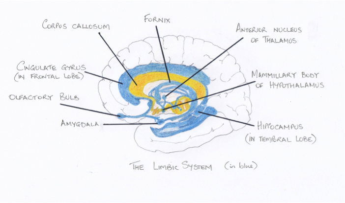
© Dr Patricia Logan 2017: The limbic system, adapted from McNaught & Callander, 1997
|
Located on the medial region of each cerebral hemisphere and diencephalon is the limbic system. It consists of the amygdaloid body, cingulate gyrus, and the hippocampus, and is our emotional brain. The amygdaloid body and the cingulate nucleus appear to be the most important structures involved in emotion with the former structure responding to perceived threats and the latter in the expression of emotion. The limbic system is believed to be the part of the brain that triggers emotional responses to smells. Connections between the limbic system and higher brain centres allows integration of external stimuli, with responses being relayed through the hypothalamus. Interaction between the frontal lobes and the limbic system allows us to have conscious input into emotional responses and as such respond appropriately.
3 The Peripheral Nervous System (PNS)
The peripheral NS is divided into the sensory and motor divisions, which is further subdivided into somatic and autonomic nervous systems.
The Somatic Nervous System
The somatic nervous system stimulates skeletal muscle in response to sensory stimuli. Sensory receptors respond to changes in their environment and relay that information to the CNS. There are several different types of receptors based on the type of stimuli detected, location of the receptor, and the structure of the receptor, and are classified as mechanoreceptors, thermoreceptors, photoreceptors, chemoreceptors, and nociceptors. Most sensory receptors are found throughout the body and consist of modified dendritic endings which receive information. Many can respond to more than one type of stimuli.
In order for one to sense the external environment several processes must occur. Initially a sensory receptor must be excited by a stimulus. For example, a photoreceptor would be excited by light, but not by touch. An action potential is then generated. Ascending sensory pathways generally consist of a chain of three neurons; fist, second and third order neurons, which ultimately take the sensory information to the cerebral cortex, where it is interpreted. Photoreceptors will always send information to the visual cortex while sound will always send impulses to the auditory cortex.
Once the brain has integrated incoming information from afferent sensory nerves, efferent motor nerves carry information away from the CNS to generate a response. The motor neuron cell bodies are located in the CNS, and have long myelinated axons that travel to the skeletal muscles they innervate. The terminals of somatic motor fibres form complex neuromuscular junctions. At the axon terminal, ACh is released which diffuses across the synaptic cleft and binds to chemical receptors opening ligand-gated ion channels, allowing the movement of sodium and potassium into and out of the muscle cell respectively. This ultimately leads to an action potential, an excitatory signal, which results in the muscle fibre contracting.
The Autonomic Nervous System (ANS)
|
|
|
The ANS innervates cardiac muscle, visceral smooth muscle, and glands. This part of the nervous system has two arms, the sympathetic and the parasympathetic. The sympathetic (stress) system activating a ‘fight or flight’ response for the body and the parasympathetic (peace) system activating ‘rest and digest’ responses in the body. Both arms usually innervate the same organs, having an opposing effect on that organ.
Innervation of autonomic motor endings in visceral smooth muscle, and glands is less complex than that of skeletal muscle. Autonomic motor axons branch repeatedly forming synapses with their respective cells. Either ACh (parasympathetic fibres) or noradrenaline (most sympathetic fibres), is released into the autonomic synaptic cleft, which acts indirectly on the target smooth muscle, or gland, resulting in either an inhibitory signal or an excitatory signal.
Neurotransmitters in the ANS
There are two critical neurotransmitters for the ANS; Acetylcholine (ACh) and the adrenergic neurotransmitters, adrenaline and the closely chemically related noradrenaline. Both divisions of the ANS use ACh for the first neuron to neuron link. The ACh binds to nicotinic receptors (named because they interact with the chemical nicotine, found also in tobacco). The second neuron in this two-step chain differs in neurotransmitter use between the sympathetic and parasympathetic division. The parasympathetic division (‘peace’ or ‘rest and digest’) division uses ACh again, but this time activating muscarinic receptors. As you probably know, the sympathetic (‘stress’ or ‘fight and flight’ division) uses adrenaline and the chemically similar noradrenaline.
4 The Reflex Arc
A reflex arc is activated in the periphery (e.g. a hand placed onto hot surface). A message is sent along a neuron to the integration centre in the spinal cord. It is processed at the level of the spinal cord (saving the time that it would take to send the message all the way to the brain). Then effector (action generating neurons) from the spinal cord carry a ‘response’ message to an effector organ (like skeletal muscle) to active a protective response – like muscle contraction to cause hand withdrawal.
5 Pain
Pain is defined by the International Association for the Study of Pain as “An unpleasant sensory and emotional experience associated with actual or potential tissue damage or described in terms of such damage.”
Activation of various pressure, tissue damage and inflammation (stretch) receptors located in the body can trigger a sensation of pain. This sensory information, like much other sensory information, is sent to the somatosensory components of the cerebral cortex. These receptors use nerve pathways that travel though brain centres responsible for awareness (which is why pain keeps people awake). Pain pathways are poorly mapped onto the cortical surface, which is why pain location is often poor and why we can get referred pain.
Pain is a major issue clinically.
Unlike most other sensations in the body, pain also has an element of perception; a subjective interpretation of the pain stimulus. We understand that pain is strongly influenced by existing patterns of brain activation, associated with cultural and personal training as well as factors such as age and anxiety. This is why pain sensation, severity and control varies from person to person.
One chemical group that is important for pain transmission is the opiate group. Opiates (those made in the body and those administered pharmacologically) reduce neurotransmitter release from pain neurons thereby limiting the transmission of messages from those neurons to the central nervous system. We ‘feel’ pain in our central nervous system (not in our body – as all sensation is ‘understood’ in our central nervous system), so if the ‘pain message’ isn’t transmitted as well, we become less ‘aware’ of the pain. This is the basis of the use of drugs like morphine.
Bibilography
Tortora, Derrickson, Burkettt, Dye, Cooke, Divers, McKean, Mellifont, Samalia & Peoples (2016). Principles of Anatomy and Physiology (1st Asia-Pacific Edition). Milton, QLD: Wiley
Seeley, Stephens & Tate (2000). Anatomy & Physiology (5th Edn). USA: McGraw-Hill
McNaught & Mackenna (1997). Illustrated Physiology. (6th Edition). New York, USA: Churchill-Livingstone
Seeley, Stephens & Tate (2000). Anatomy & Physiology (5th Edn). USA: McGraw-Hill
McNaught & Mackenna (1997). Illustrated Physiology. (6th Edition). New York, USA: Churchill-Livingstone