Learn more about Cells
In this module, you will learn more about
- The basic structure of a cell
- The internal structure of human cells: the organelles
- Transport across the cell membrane
Learn even more: See Chemistry and Introduction to Pharmacology
1 The basic structure of a cell
The cell is the basic unit of every living organism and human cells have a complex internal structure in which functions are undertaken by discrete parts.
https://en.wikipedia.org/wiki/Space_food
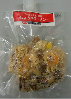
The picture is actually a sealed pack of a meal prepared for astronauts. It is a good way to think of cells since each one is a sealed-sack of components.
The major difference is that in the case of the cells usually the bulk of what is inside is water, so maybe think of a cell as a "soup-in-the-bag". The "soup" is call the cytoplasm or if someone is referring specifically to the water component: the cytosol.
The sack/or bag of the cell is the plasma membrane.
The plasma membrane
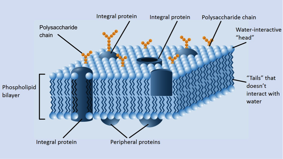
@Den.the.Grate depositphotos.com
The basic structure of the plasma membrane is shown in the diagram above.
To interpret the image, imagine a piece of the plasma membrane of a cell has been cut out and set aside so you can see both what’s on the outside and what’s in the inside.
The key features are:
To interpret the image, imagine a piece of the plasma membrane of a cell has been cut out and set aside so you can see both what’s on the outside and what’s in the inside.
The key features are:
- The bulk of the plasma membrane is a double layer (“bilayer”) of phospholipid molecules packed together with the water-interacting ends (the “head” of each molecule) in contact with the water inside or outside the cell and the other parts (the two “tails” of each molecule) that don’t interact with water turned inward
- Molecules of cholesterol are interspersed throughout the phospholipid bilayer although these aren’t shown in this diagram. They are interspersed amongst the tails of the phospholipids
- Protein molecules are found embedded in the phospholipid bilayer (“integral proteins") or a closely associated with the inner-facing or outer-facing side of the plasma membrane (“peripheral proteins")
- Polysaccharide chains may be found either attached to lipids or proteins
2 The internal structure of human cells: the organelles
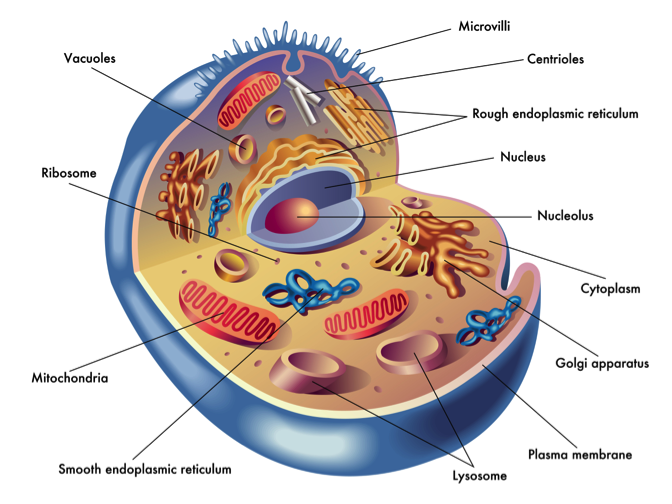
© rob3000 depositphotos.com
With the exception of bacteria, the internal anatomy of the cells of all other organisms is very complex.
You can see that in the diagram above.
So, sticking with the soup-in-bag analogy of a cell, it’s actually a chunky soup.
The chunks are the various individual and specialised functional units of the cell and they are called organelles. Which is a very appropriate name for them since it means “little organs" and these are very small structures which serve as the cell’s “organs”. Here is a short description of functions of each of the organelles found in human cells.
- Centrioles: What the diagram doesn’t show is the internal framework of proteins which give the cell its shape. This is called the cytoskeleton (literally the skeleton of the cell). The centrioles are part of this and are involved in coordination of the processes involved when one cell divides into two.
- Endoplasmic reticulum (ER): A network (“reticulum”) of membrane-walled interconnected channels that runs through the inside of the cell (“endoplasmic”). Either rough ER with ribosomes attached to it, hence a site for protein synthesis. It’s called rough because it is covered in ribosomes. Or, smooth ER which serves different roles in different cells including: calcium storage and detoxification
- Golgi apparatus/Golgi complex/Golgi body: Named after its discoverer Camillo Golgi. This is a stack of membrane walled sacks that collect proteins produced by the cells=>chemically modify them=>send them off to where the proteins are needed (e.g. for export from the cell or as lysosomes)
- Lysosomes: These are membrane-walled sacks that can contain free radicals, acids and destructive enzymes. When something is taken into a lysosome (like a non-functional organelle or an ingested bacteria) it is destroyed. Having all this occur inside the lysosome means the rest of the cell does not get damaged
- Mitochondria: One mitochondrion, many mitochondria. The major function of these is as the principal site for the synthesis of the cellular energy compound adenosine triphosphate (ATP)
- Nucleus: Contains the genes collected in molecules of DNA as well as mechanism of copying the information of the genes to be used in making proteins
- Nucleolus: Production of ribosomes
- Ribosomes: Sites of protein manufacture (“protein synthesis’)
- Vacuoles and vesicles: These are membrane-walled fluid filled sacks. For example when proteins leave the Golgi, they leave packaged in vesicles. Vesicles can fuse to make a bigger vacuole
3 Transport across the cell membrane
Movement of things in and out cells means crossing the plasma membrane and exactly how this happens will vary between different things.
http://www.kitchenkonfidence.com/2012/08/chicken-stock-recipe
|

https://pixabay.com/en/morning-tea-tea-bag-herbs-glass-750902/
|
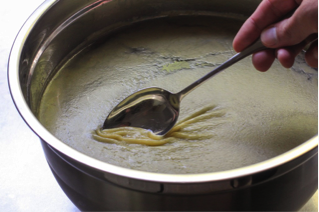
The pictures of congealed fat on the top of chicken stock and a cup of tea using a tea bag are to help you understand about things being transported across the cell membrane.
- Firstly recall that most of the plasma membrane is a layer of fat. You can imagine the plasma membrane as a layer of fat surrounding the watery cytoplasm. This layer of fat will be a barrier to the transport of materials in or out of cells
- However it is not a complete barrier. Instead it is described as being semi-permeable some things have the right chemical properties to cross the plasma membrane.
Simple diffusion
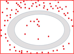
Small molecules without any electrical charge that can be dissolved in fat will easily cross the plasma membrane as long as there is a concentration gradient. Imagine this is cell and we are looking at the movement of molecules of a single chemical. Clearly the concentration of the chemical is high on the outside of the cell and low on the inside of the cell. This means there is a concentration gradient on either side of the plasma membrane. In simple diffusion, if the molecules can cross the plasma membrane, there will be preferential movement from a region of high concentration to low concentration. Oxygen crosses the plasma membrane by simple diffusion.
Facilitated diffusion
,As a nurse you are a facilitator, you will be the means of helping to patient perform tasks. Without you the task may prove too difficult for the patient or take far too long.
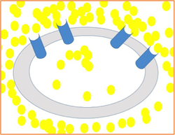
For molecules that can’t cross the plasma membrane because they are too large and/or have an electrical charge: what’s needed is a facilitator. In this case it is a protein embedded in the plasma membrane which essentially ferries the molecule across. This is facilitated diffusion.
The protein responsible for the facilitated diffusion of a particular molecule will be specific for that molecule. So, for glucose to be transported across the plasma membrane there must be specific glucose transport proteins embedded in that plasma membrane.
While the amount of movement that occurs with simple diffusion is dependent on the concentration gradient, facilitated diffusion relies BOTH on a concentration gradient AND the number of transport proteins present. This is just like traffic movement across a bridge dependent on how many lanes are available.
Osmosis
Water is a small molecule and it also has no charge. So water molecules can move across the plasma membrane. This process is greatly assisted by the presence of specific water-transporting protein channels running through the plasma membrane.
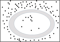
Here’s another situation with a concentration gradient. In this case ability of the molecule to cross the plasma membrane is restricted. So to “balance things out” water will cross the plasma membrane instead. Since the outside of the cell is more concentrated than the inside of the cell: the preferential movement of the water will be to outside of the cell.
Osmosis is a critical process in the transport of water from the lumen of the gastrointestinal tract (GIT) and ultimately into the GIT blood stream. Osmosis is critical in the process where the kidneys reduce the amount of water lost in urine if someone is dehydrated.
Other forms of movement across the plasma membrane:
- For normal cell function, the cytosol must have a high potassium concentration and a low sodium concentration. This is achieved by what is called a solute pump, in this particular case it’s an embedded protein called the Na+K+-ATPase. This pump moves potassium into cells and sodium out of cells even though this is against an existing normal concentration gradient. In order to do this, chemical energy (ATP) must be consumed.
- Materials transported in vesicles can be delivered to the plasma-membrane for export from the cell. This process is called exocytosis (“outside the cell”)
- Parts of the plasma membrane can extend out and trap small volumes of the fluid (pinocytosis) or particles (like a bacteria or a bit of dead cell, and the process is known as phagocytosis). The portion of plasma membrane buds off as a vesicle and so delivers either the fluid or the particle to the inside of the cell. Both cases are examples endocytosis (“inside the cell”)
- In some cases it is the binding of a molecule to a specific protein on the outer surface of the plasma membrane that is a trigger for endocytosis.